Heart Anatomy Draw
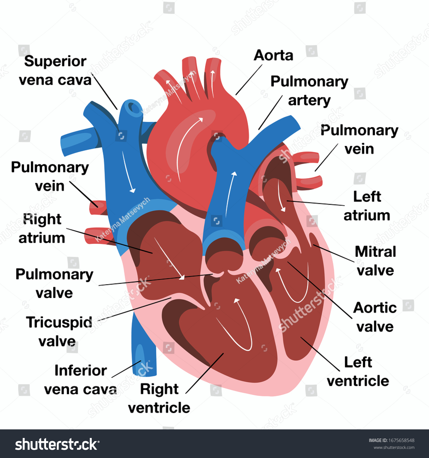
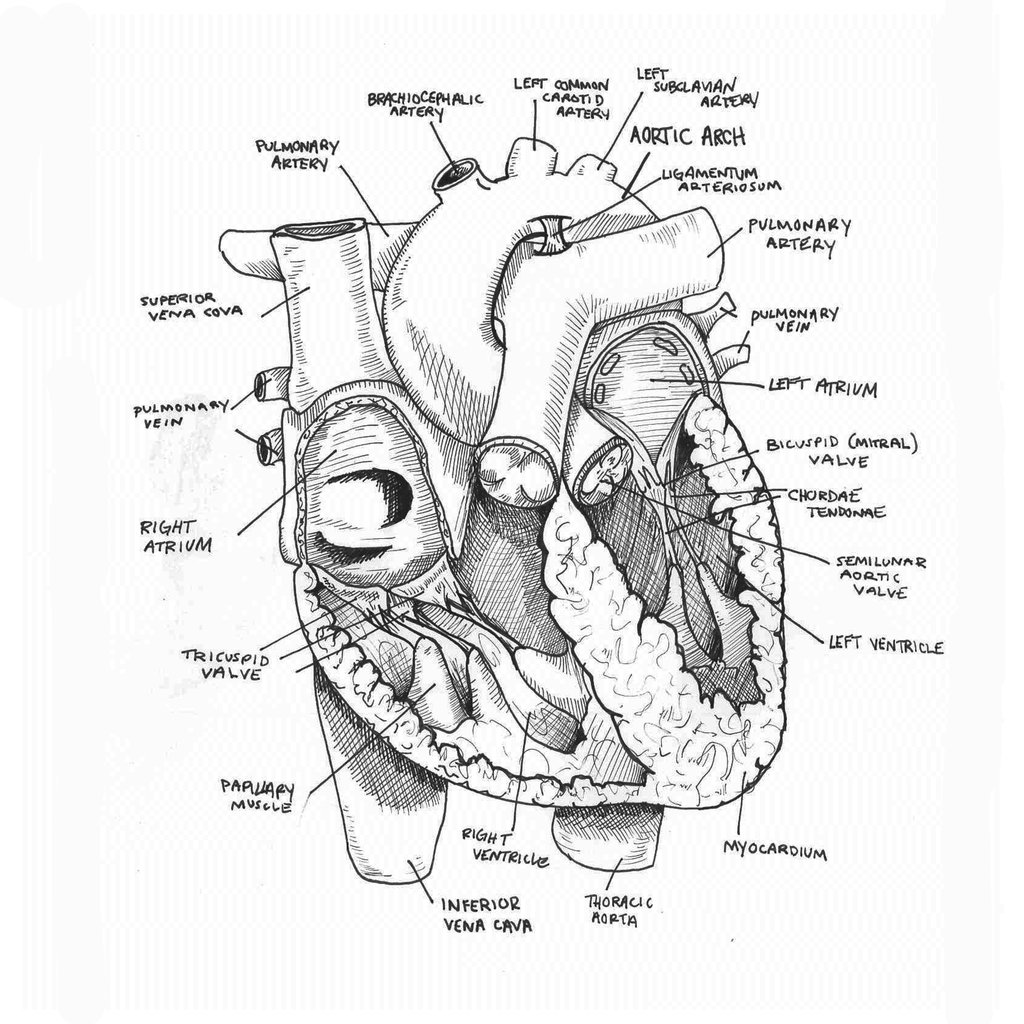
Heart Anatomy Draw - The inferior tip of the heart, known as the apex, rests just superior to the diaphragm. Web the surfaces and borders of the heart. Web in this interactive, you can label parts of the human heart. Understanding its basic anatomy is crucial to understanding how it functions. You have two chambers on the top (atrium, plural atria) and two on the bottom (ventricles), one on each side of your heart. Web with this easy human heart drawing ideas, you can learn how to draw a human heart easily. If you want to redo an answer, click on the box and the answer will go back to the top so you can move it to another box. Web this interactive atlas of human heart anatomy is based on medical illustrations and cadaver photography. Web the heart is shaped as a quadrangular pyramid, and orientated as if the pyramid has fallen onto one of its sides so that its base faces the posterior thoracic wall, and its apex is pointed toward the anterior thoracic wall. Give you the option to test yourself with english or latin terminology. Dr matt & dr mike. Drawing a human heart is easier than you may think. Web this interactive atlas of human heart anatomy is based on medical illustrations and cadaver photography. Web these clever heart anatomy quizzes will. Bones of the pelvis the sacroiliac joint. Sketch out a basic outline of the heart, using our tutorial as a guide. Web the intricate anatomy of the heart can be challenging to grasp, and so i hope you find this tool to be helpful in visualizing the cardiac system. On its superior end, the base of the heart is attached to the aorta, pulmonary arteries and veins, and the vena cava. The innermost layer is thin and smooth. This method breaks down complex structures into simple shapes, making the drawing process approachable and enjoyable for artists at any skill level. A drawing of the anatomy of the opened normal heart, with english labels. If you want to redo an answer, click on the box and the answer will go back to the top so you can move it to another box. There is also a murmurs tab that includes some of the more common murmurs we come across. Web the. On its superior end, the base of the heart is attached to the aorta, pulmonary arteries and veins, and the vena cava. Human heart drawing step by step guide. Web in this interactive, you can label parts of the human heart. Adapt to your knowledge, repeating questions that you got wrong. Sketch out a basic outline of the heart, using. Web drawing the human heart can be a challenging yet rewarding artistic endeavor. Web the heart is shaped as a quadrangular pyramid, and orientated as if the pyramid has fallen onto one of its sides so that its base faces the posterior thoracic wall, and its apex is pointed toward the anterior thoracic wall. Then, fill in the base of. Sketch out a basic outline of the heart, using our tutorial as a guide. Web best way to draw and label the heart! Give you the option to test yourself with english or latin terminology. Two atria and two ventricles. We will then proceed to shape the heart, slowly refining it with our pencils into a. The valves of the heart. Give you the option to test yourself with english or latin terminology. The inferior tip of the heart, known as the apex, rests just superior to the diaphragm. Sketch out a basic outline of the heart, using our tutorial as a guide. Web function and anatomy of the heart made easy using labeled diagrams of. Web to draw an anatomical heart realistically, pay attention to the proportions and positioning of the different parts of the heart, as well as their texture and color. Web the intricate anatomy of the heart can be challenging to grasp, and so i hope you find this tool to be helpful in visualizing the cardiac system. I made this cool. Web the heart is located in the thoracic cavity medial to the lungs and posterior to the sternum. Drag and drop the text labels onto the boxes next to the heart diagram. A drawing of the anatomy of the opened normal heart, with english labels. Web to draw the internal structure of the heart, start by sketching the 2 pulmonary. If you want to redo an answer, click on the box and the answer will go back to the top so you can move it to another box. The heart is divided into four chambers: Web the intricate anatomy of the heart can be challenging to grasp, and so i hope you find this tool to be helpful in visualizing. The superior vena cava carries blood from your upper body. 48k views 1 year ago cardiovascular. Web this interactive atlas of human heart anatomy is based on medical illustrations and cadaver photography. It consists of four main chambers: The user can show or hide the anatomical labels which provide a useful tool to create illustrations perfectly adapted for teaching. The heart is divided into four chambers: Drawing a human heart is easier than you may think. Two atria and two ventricles. The inferior tip of the heart, known as the apex, rests just superior to the diaphragm. Web your heart sure does work hard, but that doesn’t mean you have to work hard to draw it! It consists of four main chambers: Blood is transported through the body via a complex network of veins. The superior vena cava carries blood from your upper body. Two atria and two ventricles. It is divided into the left and right sides by a muscular wall called the septum. Web in animals with lungs —amphibians, reptiles, birds, and mammals—the heart shows various stages of evolution from a single to a double pump that circulates blood (1) to the lungs and (2) to the body as a whole. On its superior end, the base of the heart is attached to the aorta, pulmonary arteries and veins, and the vena cava. Drag and drop the text labels onto the boxes next to the heart diagram. Web the intricate anatomy of the heart can be challenging to grasp, and so i hope you find this tool to be helpful in visualizing the cardiac system. Web to draw the internal structure of the heart, start by sketching the 2 pulmonary veins to the lower left of the aorta and the bottom of the inferior vena cava slightly to the right of that. There is also a murmurs tab that includes some of the more common murmurs we come across. Adapt to your knowledge, repeating questions that you got wrong. From the openstax anatomy and physiology book. Web the heart is located in the thoracic cavity medial to the lungs and posterior to the sternum. Sketch out a basic outline of the heart, using our tutorial as a guide. Web your heart has four separate chambers.
anatomically correct human heart by NIku Arbabi embroidery

Diagrams of Human Heart Diagram Link Heart diagram, Human heart

How to Draw the Internal Structure of the Heart (with Pictures)
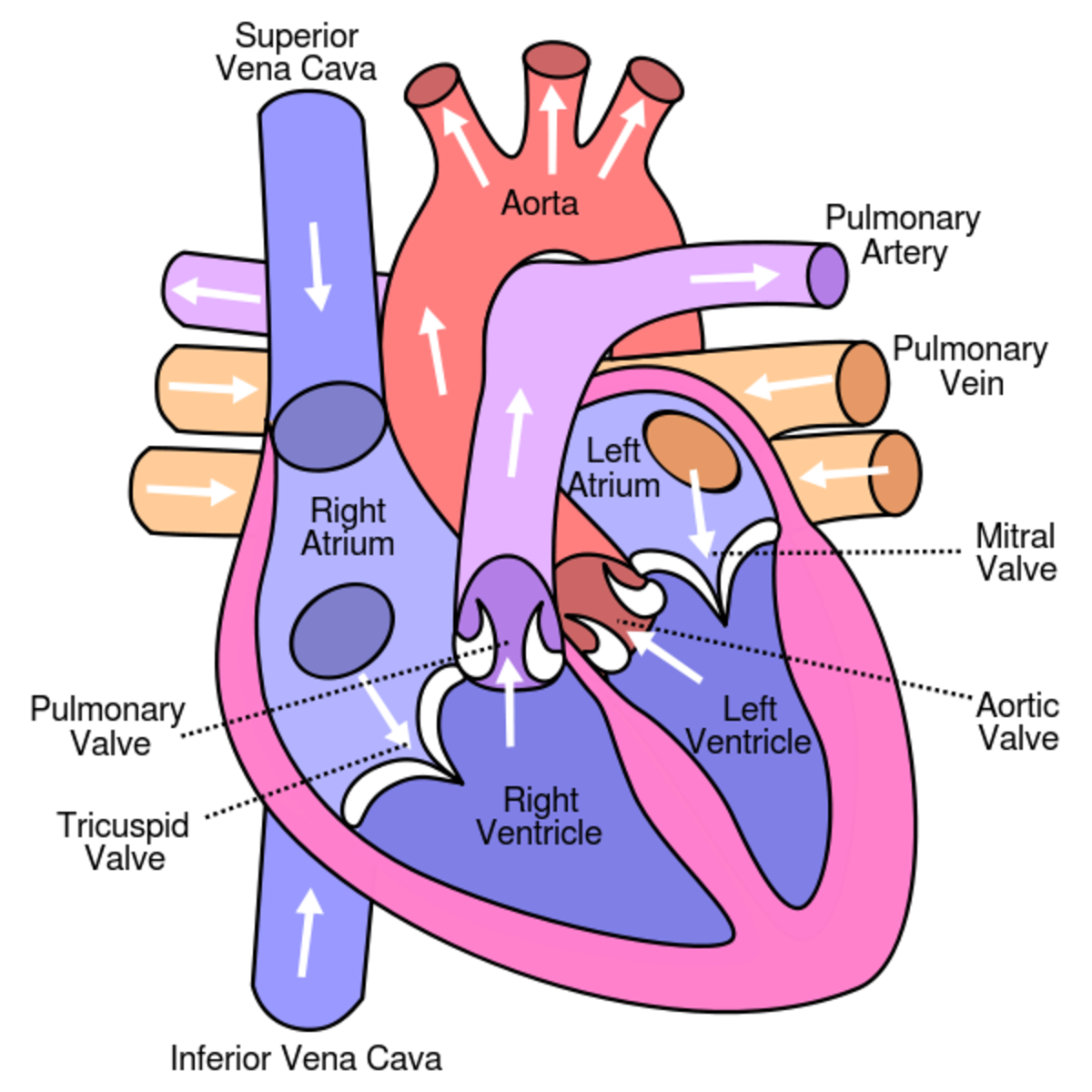
Learn About the Heart and Circulatory System for Kids hubpages
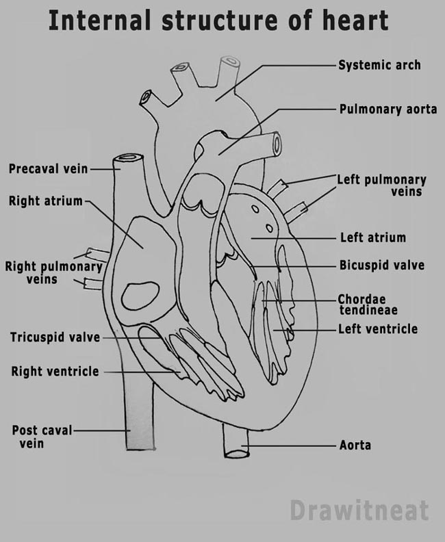
DRAW IT NEAT How to draw human heart labeled

How to Draw the Internal Structure of the Heart 13 Steps
Hand Drawn Illustration Human Heart Anatomy Stock Vector (Royalty Free

Human Heart Drawing & Labelling YouTube

Human heart anatomy. Vector diagram in 2021 Heart anatomy, Human
Anatomical Drawing Heart at GetDrawings Free download
Web Drawing The Human Heart Can Be A Challenging Yet Rewarding Artistic Endeavor.
Web Best Way To Draw And Label The Heart!
Web The Surfaces And Borders Of The Heart.
The Innermost Layer Is Thin And Smooth.
Related Post: